The appearance of fine red or purple clusters on the legs, commonly known as spider veins or telangiectasias, is a concern that extends beyond cosmetic appearance. For many patients, these visible vascular networks represent a barrier to confidence and a sign of underlying venous pressure. While often asymptomatic, their presence may indicate early stages of chronic venous insufficiency (CVI).
In the realm of leg aesthetics, sclerotherapy for spider veins remains the undisputed gold standard. Despite the rise of transdermal lasers, sclerotherapy offers a level of precision and “root-cause” treatment that light-based therapies often struggle to match.
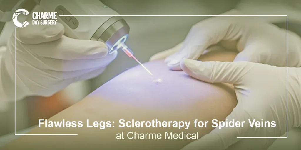
Sclerotherapy for Spider Veins: Procedure & Benefits
Sclerotherapy is a minimally invasive medical procedure designed to eliminate “thread veins” and small varicose veins. The process involves targeted injection of a sclerosing agent directly into the affected blood vessel.
Clinical Benefits of Sclerotherapy
The primary advantage of sclerotherapy is its ability to treat the “feeder veins” that are often invisible to the naked eye but are responsible for the visible spider veins on the surface.
- Minimal Downtime: Unlike surgical Varicose Veins Treatment, sclerotherapy is an “office-based” procedure requiring no anesthesia.
- High Success Rate: Modern sclerosants have a high safety profile and effectively clear 50% to 80% of treated veins per session.
- Improved Leg Aesthetics: Beyond the medical benefits, the psychological impact of having “clear legs” significantly improves patient quality of life and comfort in social or athletic settings.
Spider Veins vs. Varicose Veins: What’s the Difference?
In clinical phlebology, we use the CEAP classification to categorize venous disease. Understanding where a patient falls on this scale is vital for choosing the right intervention.
Spider Veins (Telangiectasias):
These are classified as CEAP C1 lesions. They are small (less than 1 mm in diameter), superficial, and usually appear as sunburst or “web-like” patterns. They reside in the dermis and do not typically cause significant swelling.
Reticular Veins (Feeder Veins):
Often called “feeder veins,” these are bluish-green veins (1–3mm) that sit slightly deeper. They often supply the high pressure that creates spider veins.
Varicose Veins (CEAP C2 and Above):
These are CEAP C2 or higher. They are large, bulging, and tortuous (greater than 3mm). While sclerotherapy can treat some varicose veins (particularly via foam), they often require more robust interventions like EVLA laser or radiofrequency ablation.
By identifying the presence of reticular “feeders” via transillumination or ultrasound, a skin specialist ensures that thread vein removal is long-lasting rather than temporary.
Read also about: Varicose Veins Treatment
How Sclerotherapy Works (Injection Technique)
The mechanism of sclerotherapy is rooted in controlled “endothelial injury.” The sclerosing agent, most commonly Polidocanol (standard in Europe/Spain) or Sodium Tetradecyl Sulfate, acts as a surfactant that disrupts endothelial cell integrity.
The Biological Process
- Injection: Using an ultra-fine needle, the specialist performs spider veins injections into the vessel.
- Endothelial Damage: The solution irritates the delicate inner lining (endothelium) of the vein.
- Vasospasm and Thrombosis: The vein immediately spasms and the walls stick together. A small, controlled clot forms, which effectively permanently closes the vessel.
- Fibrosis and Reabsorption: Over the following weeks, the body’s inflammatory response replaces the dead vein with fibrous tissue. Eventually, the immune system’s macrophages reabsorb the tissue, and the vein disappears from sight.
Foam vs. Liquid Sclerotherapy
The choice between liquid and foam is determined by the size and depth of the target vessel.
Liquid Sclerotherapy
Liquid is the traditional medium for the smallest telangiectasias. Because liquid flows easily, it can fill the tiny “capillary” networks of spider veins. However, in larger vessels, liquid can become too diluted by blood, reducing its effectiveness.
Foam Sclerotherapy
Foam sclerotherapy (often created using the “Tessari Technique,” widely documented in Italian and French vascular journals) involves mixing the liquid sclerosant with air or a physiological gas (like O2/CO2).
- Increased Surface Area: The foam consistency allows the chemical to displace blood rather than mix with it, ensuring near-complete contact with the vein wall.
- Enhanced Visibility: Foam is highly visible on ultrasound, making it the preferred choice for treating deeper “feeder” veins or recurrent varicose veins.
Who is a Candidate for This Treatment?
While most individuals seeking cosmetic vein treatment are eligible, a clinical consultation is mandatory to rule out contraindications.
Ideal Candidates:
Non-smokers with localized spider veins and no significant underlying venous reflux.
Contraindications:
- A history of Deep Vein Thrombosis (DVT) or pulmonary embolism.
- Acute skin infections in the treatment area.
- Known allergy to sclerosing agents.
- For foam sclerotherapy: A known “Patent Foramen Ovale” (PFO), which is a small hole in the heart, as the foam bubbles could theoretically enter the arterial circulation.
A thorough medical history and, in many cases, a Duplex Ultrasound are used to ensure that the spider veins are not a symptom of “Deep Vein Reflux,” which would require a different treatment path.
Post-Treatment Care (Compression Stockings)
The success of sclerotherapy for spider veins depends equally on proper technique and post-treatment care. The most critical component of recovery is the use of medical-grade compression stockings (usually Class II, 20–30 mmHg).
Why Compression is Essential:
- Vessel Apposition: Compression keeps the treated vein walls pressed together, ensuring they “seal” permanently.
- Prevention of Trapped Blood: By minimizing the amount of blood that can re-enter the treated vein, compression reduces the risk of “hemosiderin staining”, the brownish pigmentation that can occur if iron from the blood leaks into the skin during healing.
- Reduced Side Effects: It minimizes the risk of “matting” (the growth of tiny new capillaries) and reduces post-procedure inflammation.
Patients are typically advised to wear stockings for 3 to 7 days continuously (depending on the vessel size) and avoid high-impact exercise or hot baths during the initial healing phase.
FAQ
Is sclerotherapy painful?
The procedure is remarkably well-tolerated. Most patients describe the spider veins injections as a tiny “mosquito bite.” The sclerosant itself (especially Polidocanol) has mild anesthetic properties, so any stinging sensation usually dissipates within minutes.
How many sessions do I need for spider veins?
While some veins vanish after one session, most patients require 2 to 4 sessions for optimal results. These sessions are typically spaced 4 to 6 weeks apart to allow the body time to clear the treated vessels.
Can I exercise after sclerotherapy?
Walking is highly encouraged immediately after the procedure. However, “high-impact” activities (running, heavy weightlifting, or high-intensity interval training) should be avoided for 5–7 days to prevent the treated veins from reopening under high pressure.